


الاخبار


اخبار الساحة الاسلامية

أخبار العتبة العلوية المقدسة
أخبار العتبة الحسينية المقدسة
أخبار العتبة الكاظمية المقدسة
أخبار العتبة العسكرية المقدسة
أخبار العتبة العباسية المقدسة
أخبار العلوم و التكنولوجيا
الاخبار الصحية
الاخبار الاقتصادية

لمن تجاوز الستين.. عليك بشرب 4 أكواب من القهوة يوميا ؟
 المصدر:
skynewsarabia.com
المصدر:
skynewsarabia.com
 02:18 صباحاً
 2025-07-01
 75
75


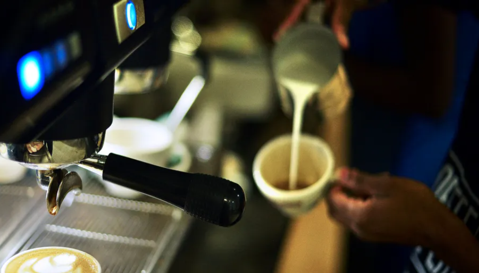
كشفت دراسة، منشورة حديثا في المجلة الأوروبية للتغذية، أن شرب 4 إلى 6 أكواب من القهوة يوميا يُخفض الهشاشة ويحسن من النشاط الجسدي والعقلي لدى كبار السن.
وذكر تقرير لصحيفة "التليغراف" البريطانية أن الدراسة أُجريت على مدى 4 سنوات، وشملت 1161 شخصا يبلغون من العمر 55 سنة فأكثر، ضمن برنامج الشيخوخة الطولية في أمستردام، وهي أول دراسة من نوعها تحلل العلاقة بين القهوة والهشاشة.
واعتمد البحث على تقييم الصحة الجسدية للمشاركين، بالتركيز على فقدان الوزن، والضعف، والإرهاق، وبطء المشي، وانخفاض النشاط البدني.
وأظهرت بيانات الأبحاث أن شرب القهوة يحسن الإدراك ويعزز نشاط العضلات، ويقي من الهشاشة. وأرجع الباحثون ذلك إلى مكوناتها المفيدة للجسم.
فالكافيين، المكون الرئيسي للقهوة، يساعد في التقليل من التعب وزيادة اليقظة، إضافة إلى تسهيل حركة العضلات وتيسير القدرة على التنقل.
كما أن البوليفينولات، تحارب الالتهاب والأكسدة، وتقي من وهن العضلات، وتقلل من التورم، وتدعم الوظائف العامة للجسم.
أما مركب تريغونيلين، غير المعروف، فيحافظ على الصحة الإدراكية ويحسن الذاكرة.
غير أن الخبراء، أكدوا أن الإفراط في شرب القهوة له نتائج عكسية.
وتقول ديل ستانفورد، أخصائية التغذية في مؤسسة القلب البريطانية: "إن استهلاك كوبين أو ثلاثة من القهوة يوميا قد يرتبط بانخفاض خطر الإصابة بأمراض القلب مقارنة بعدم شرب القهوة. لكن شرب أكثر من أربعة أو خمسة أكواب يوميا قد يرفع معدل استهلاك الكافيين إلى ما فوق الحد اليومي الموصى به، وهو 400 ملغ تقريبا (أي ما يعادل أربعة إلى خمسة أكواب)".
ويمكن أن يسبب الإفراط في شرب القهوة في ارتفاع ضغط الدم، وتسارع ضربات القلب، والقلق، والتوتر، والغثيان، والصداع، واضطرابات النوم.
هل تحل القهوة محل التمارين الرياضية؟
ذكر التقرير ذاته أن القهوة هي مكمل، ولا يجب أن تحل محل التمارين الرياضية، لأن النشاط البدني المنتظم يحسن من القوة والقدرة على التحمل، ويعزز قدرة الجسم على أداء وظائفه، بينما الكافيين مجرد أداة داعمة تؤخر الشعور بالتعب.
وينصح الأطباء بممارسة النشاط البدني بشكل منتظم، واتباع نظام غذائي متوازن غني بالأطعمة المضادة للأكسدة مثل القهوة.
أطعمة تكافح الهشاشة
نصح التقرير بتناول التوت، والشوكولاتة الداكنة، وزيت الزيتون، والشاي الأخضر، والكثير من الخضروات. إضافة إلى الأطعمة الغنية بالبروتين كالبيض، ومشتقات الحليب، والبقوليات، والسمك، واللحوم الخالية من الدهون.
اخبار ذات صلة
 (نوافذ).. إصدار أدبي يوثق القصص الفائزة في مسابقة الإمام العسكري (عليه السلام)
 قسم الشؤون الفكرية يصدر مجموعة قصصية بعنوان (قلوب بلا مأوى)
 قسم الشؤون الفكرية يصدر مجموعة قصصية بعنوان (قلوب بلا مأوى)


