آخر المواضيع المضافة

 النبات
الحيوان
الأحياء المجهرية
علم الأمراض
التقانة الإحيائية
التقنية الحيوية المكروبية
التقنية الحياتية النانوية
علم الأجنة
الأحياء الجزيئي
علم وظائف الأعضاء
الغدد
المضادات الحيوية
النبات
الحيوان
الأحياء المجهرية
علم الأمراض
التقانة الإحيائية
التقنية الحيوية المكروبية
التقنية الحياتية النانوية
علم الأجنة
الأحياء الجزيئي
علم وظائف الأعضاء
الغدد
المضادات الحيوية| Cell Death |


|
|
Read More
Date: 17-5-2021
Date: 8-4-2021
Date: 21-11-2015
|
Cell Death
It is not initially obvious why cell death should be considered important. The death of single-celled organisms, such as amoeba or bacteria, has no apparent advantage for the individual cell. However, in multicellular organisms damage to an individual cell can have repercussions for the whole animal. It therefore makes biological sense to dispose of this cell and replace it with a healthy one. Cells become damaged in a variety of different ways, but for long-lived, multicellular animals, the greatest risk is from a damaged cell that has acquired an oncogenic mutation leading to unrestricted clonal growth. To minimize this risk, several pathways have evolved that limit the expansion of somatic cells, one of which is cell death.
Tissue size is restricted by several different methods, not all of which actually require the physical loss of the cell. The problem facing multicellular organisms is how to maintain the capacity to divide and replace damaged cells while minimizing the risk of mutation leading to enhanced growth potential. One way around this problem is to make tissues post-replicative, as occurs in the central nervous system (CNS). All the neurons within the CNS are produced during embryogenesis and are then maintained throughout the life span of the organism. For some tissues, however, such an approach is not practical due to persistent physical damage to the cells, such as occurs in the epithelial lining of the gut or skin. Here, cells are continuously produced by a select number of proliferating stem cells. The daughter cells they produce move upward, away from the basement membrane, terminally differentiate, and are eventually shed. Because these cells are continuously replaced, any damaged cell should be automatically eliminated. Tissue size is also restricted by the need for vascularization. If there is no blood supply, only a small proportion of cells can be maintained by the diffusion of solutes, which is one of the reasons why many solid tumors have central necrotic zones.
For tissues that require a significant number of cells in cycle, the risk of mutation is minimized by both senescence and cell death. Senescence is a pathway that is invoked once cells have undergone a specific number of doublings, causing them to arrest permanently. Cells in this senescent state are unable to reenter the cell cycle (1). Thus, a cell that does acquire a mutation allowing constant proliferation is limited in its capacity to divide, restricting clonal outgrowth. Finally, cells can be triggered to die when no longer required, or when damaged. This “programmed cell death” is essential for the maintenance of tissue homeostasis and is invoked in disparate situations including DNA damage, absence of survival signals, and oncogene activation (2, 3). Thus, multicellular organisms are protected against somatic mutation by a number of independent mechanisms that act in concert to limit the potential for neoplastic transformation.
1. The Meaning of Cell Death
The concept that cell death is fundamentally important for restricting cell population expansion took some time to be accepted for several reasons. Dead cells are not obvious in healthy tissues, whereas they are abundant in areas of damaged tissue, such as ischemic heart tissue resulting from myocardial infarction, allowing dead cells to be fundamentally associated with disease. Only during embryogenesis are dying cells seen in abundance, but these were generally regarded as cells deleted as a result of overproduction during development. In addition, death is seen as a bad outcome in human terms, a misconception that has allowed human anthropomorphic confusions to outweigh scientific evidence. More recently, however, several of these objections have been overruled, allowing cell death to become an accepted, essential daily process.
Early investigators of both invertebrate and vertebrate development observed that developmental cell deaths occurred in response to several biological cues and could be suppressed by inhibitors of both protein and RNA synthesis (4, 5), suggesting a requirement for macromolecular synthesis (6, 7). Moreover, they made the important connection that these cell deaths were an essential part of the developmental program of the organism concerned; hence the term “programmed cell death” (PCD) (8). It is now possible in less complex invertebrate models, such as the nematode Caenorhabditis elegans, to map both the fate of all individual cells within the developing organism and the genes that dictate these fates (9).
For vertebrates, the importance of cell death was not appreciated until the detailed characterization of the form of cell death termed apoptosis (10). The programmed cell deaths observed during development are identical to apoptotic cells in mammalian tissues, suggesting that a regulated form of cell death has been conserved throughout evolution (11, 12). From these early observations, our understanding of apoptosis/PCD has grown to include a complex pathway that is both morphologically and biochemically distinct from classical necrosis or accidental cell death. Necrosis is a passive event, in which cells that become irreversibly damaged, and therefore useless, die (13, 14). This form of cell death, which requires no input from the dying cell, occurs in cells subject to physical disruption or severe toxic stress. Conversely, apoptosis/PCD describes a pathway of events in which the cell is actively involved. Due to the active nature of apoptosis, which is triggered by many physiological and toxic stimuli, this form of cell death is sometimes referred to as “cell suicide.” Overall, the importance of “active” cell death is underlined by the fact that death is the default state for all cells. Hence, all cells are programmed to die, unless signaled to survive (15).
The importance of cell death in the regulation of tissue homeostasis is paramount. The number of proliferating cells determines the cell population number, as does the number of differentiating cells and the number of dying cells. Research over the last 20 years has graphically shown how cells that have mutations which disrupt their capacity to undergo apoptosis in response to physiological stimuli are involved in the etiology of several diseases, including Alzheimer's, AIDS, and cancer (16).
Aside from the three pathways of death described above, the “death” of a cell does not always result in the loss of viability or cellular function. Within a cell population, few cells are actually proliferating, many are in a resting state outside of the cell cycle, known as GM0 phase. Certain cells can reenter the cell cycle given the appropriate stimuli, but cells that enter a permanent G0 state do not respond to these proliferation signals. Cells in the latter state are termed senescent and can complete all normal functions except division (1). Cells that escape the confines of senescence are able to proliferate continuously and, more important, the mutations that lead to the evasion of senescence are common and perhaps mandatory in tumor cells (17).
Thus, the description “cell death” encapsulates several diverse processes. This can involve the physical loss of the cells through apoptosis/PCD or, in some circumstances, necrosis. Alternatively, cell death can refer to a genetic death, where cells no longer retain the ability to replicate, but continue to survive. Which method of cell death is induced depends on several factors, including the cell's internal environment, its external environment, and its developmental history. It is not yet clear whether the genes that regulate apoptosis/PCD and senescence overlap. For example, genes such as p53 and retinoblastoma appear to be required for the regulation of apoptosis and senescence, but it is not clear whether these genes perform similar or different roles in each pathway. Indeed, cells that have evaded the first signal to senesce enter crisis, which is defined as the point at which the culture exhibits both apoptosis and proliferation. This suggests that there must be some overlap in the control of different types of cell death, and this may enable one type of cell death to be employed when another either is not suitable or is not able to be induced.
References
1. C. A. Afshari and J. C. Barrett (1996) "Molecualr genetics of in vivo cellular senescence". In Cellular Aging and Cell Death (N. J. Holbrook, G. R. Martin, and R. A. Lockshin, eds.), Wiley-Liss, New York, pp. 109–122.
2. A. J. Hale, C. A. Smith, L. C. Sutherland, V. E. Stoneman, V. Longthorne, A. C. Culhane, and G. T. Williams (1996) Apoptosis: Molecular regulation of cell death. Eur. J. Biochem. 237, 884.
3. S. J. Martin, and D. R. Green (1995) Apoptosis and cancer: The failure of controls on cell death and cell survival. Crit. Rev. Oncol. Hematol. 18, 137–153.
4. J. R. Tata (1966) Requirement for RNA and protein synthesis for induced regression of the tadpole tail in organ culture. Dev. Biol. 13, 77–94.
5. R. A. Lockshin (1969) Programmed cell death. Activation of lysis by a mechanism involving the synthesis of protein. J. Insect Physiol. 15, 1505–1516.
6. A. Glucksmann (1965) Cell death in normal development. Arch. Biol. Liege 76, 419–437.
7. P. G. Clarke and S. Clarke (1996) Nineteenth century research on naturally occurring cell death and related phenomena. Anat. Embryol. Berl. 193, 81–99.
8. R. A. Lockshin and C. M. Williams (1964) Programmed cell death. II. Endocrine potentiation of the breakdown of the intersegmental muscles of silkmoths. J. Insect Physiol., 10, 643.
9. R. E. Ellis, J. Y. Yuan, and H. R. Horvitz (1991) Mechanisms and functions of cell death. Ann. Rev. Cell Biol. 7, 663–698.
10. A. H. Wyllie, J. F. Kerr, and A. R. Currie (1972) Cellular events in the adrenal cortex following ACTH deprivation. J. Pathol. 106, ix.
11. M. D. Jacobson, M. Weil, and M. C. Raff (1997) Programmed cell death in animal development. Cell 88, 347–354.
12. A. Fraser, N. McCarthy, and G. I. Evan (1996) Biochemistry of cell-death. Curr. Opin. Neurobiol. 6, 71–80.
13. B. F. Trump, J. M. Valigorsky, J. H. Dees, W. J. Mergner, K. M. Kim, R. T. Jones, R. E. Pendergrass, J. Garbus, and R. A. Cowley (1973) Cellular change in human disease. A new method of pathological analysis. Hum. Pathol. 4, 89–109.
14. A. H. Wyllie, J. F. Kerr, and A. R. Currie (1980) Cell death: The significance of apoptosis. Int. Rev. Cytol. 68, 251–306.
15. M. C. Raff (1992) Social controls on cell survival and cell death. Nature 356, 397–400.
16.C. B. Thompson (1995) Apoptosis in the pathogenesis and treatment of disease. Science 267, 1456-1462.
17. W. E. Wright and J. W. Shay (1996) "Mechanisms of escaping senescence in human diploid cells". In Celluar Aging and Cell Death (N. J. Holbrook, G. R. Martin, and R. A. Lockshin, eds.), Wiley-Liss, New York, pp. 153–166.


|
|
|
|
لخفض ضغط الدم.. دراسة تحدد "تمارين مهمة"
|
|
|
|
|
|
|
طال انتظارها.. ميزة جديدة من "واتساب" تعزز الخصوصية
|
|
|
|
|
|
|
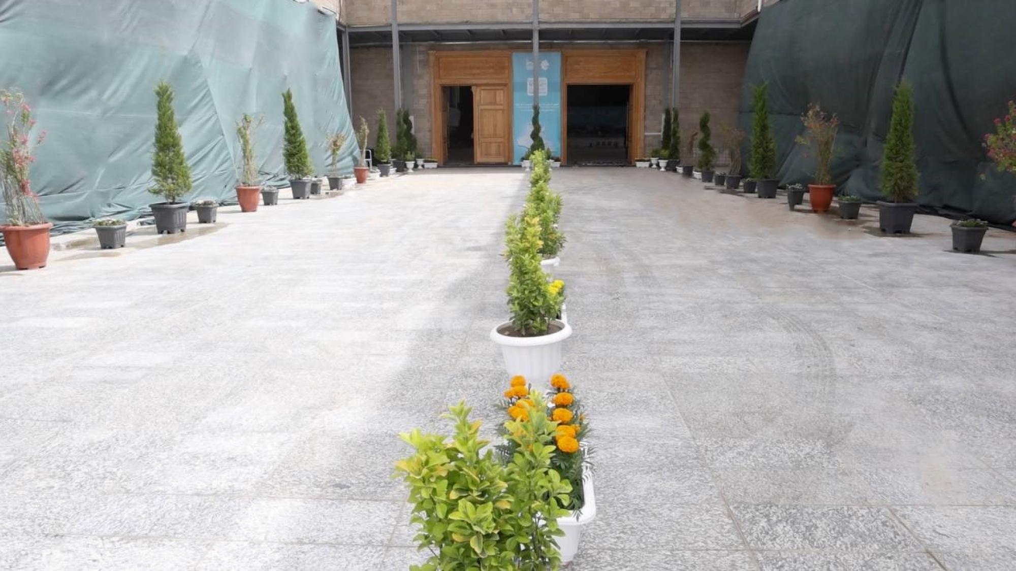
مشاتل الكفيل تزيّن مجمّع أبي الفضل العبّاس (عليه السلام) بالورد استعدادًا لحفل التخرج المركزي
|
|
|