آخر المواضيع المضافة

 النبات
الحيوان
الأحياء المجهرية
علم الأمراض
التقانة الإحيائية
التقنية الحيوية المكروبية
التقنية الحياتية النانوية
علم الأجنة
الأحياء الجزيئي
علم وظائف الأعضاء
الغدد
المضادات الحيوية
النبات
الحيوان
الأحياء المجهرية
علم الأمراض
التقانة الإحيائية
التقنية الحيوية المكروبية
التقنية الحياتية النانوية
علم الأجنة
الأحياء الجزيئي
علم وظائف الأعضاء
الغدد
المضادات الحيوية| Chemokines |


|
|
Read More
Date: 21-11-2020
Date: 30-4-2021
Date: 28-10-2020
|
Chemokines
Chemokines are small secreted proteins, generally 8 to 15 kDa in mass, that were originally identified by their ability to stimulate chemotaxis and/or activate leukocytes (lymphocytes, neutrophils, eosinophils, mast cells, monocytes, and macrophages). They are now known to be multifunctional and to be secreted by many different cell types in response to various stimuli (for reviews see Refs. 1-5). In inflamed tissues, they may induce effector functions such as the generation of an oxidative burst or the secretion, from storage granules, of proteinases (by neutrophils and monocytes), histamine (by basophils), and cytotoxic proteins (by eosinophils). Chemokines activate cell-surface receptors to promote adhesion of circulating leukocytes to the vascular endothelium and subsequently to facilitate extravasation (diapedesis) of the adherent leukocyte into adjacent tissue, the source of the chemokine. They have proinflammatory effects and can give rise to acute or chronic inflammatory responses; some have been shown to be involved in movement and activation of cells in both angiogenesis/vasculogenesis and neural pathfinding, thus implicating them in aspects of development not directly involved in the immune response.
1. Chemokine Structure and Chromosome Location
The chemokine family (at least 40 members have been identified in humans) consists of four classes distinguished by the positioning of four conserved cysteine residues near the N-terminus of the protein. These four cysteine residues define a distinct structural motif based on the two disulfide bonds (C16C3, C26C4) that they form. All the chemokines possess a central b-pleated sheet region consisting of three antiparallel beta-strands (a Greek key motif) followed in most cases by a C-terminal alpha-helix. There is evidence that the chemokines can form dimers and higher oligomers, but whether this is important for their function is controversial (2). The N-terminal sequence of the chemokines is of particular importance in determining their specificity. Alteration or removal of one or a few amino acid residues can dramatically affect their activity or target cell specificity. This provides the opportunity for the specificity of a chemokine to be altered by local factors subsequent to its secretion.
The CXC or a-class chemokines possess a single residue between the first and second cysteines. This family can be further subdivided by the presence or absence of the amino acid sequence Glu-Leu-Arg (ELR) preceding the first cysteine. This sequence is recognized by chemokine receptors on specific cell types, and thus, has functional implications. The CC or b-class chemokines have no residues between the first and second cysteines. Some members of this group have two additional cysteine residues that form a third disulfide bond (6).
Two additional chemokines have been identified that do not fit into either the CC or CXC classes. The g class is missing both the first and third cysteines. This C chemokine has lymphotactin as its prototype member. The d class possesses a CXXXC motif, and thus far is represented only by fractalkine/neurotactin. This atypical membrane-bound CXXXC chemokine has, on the C-terminal side of the b-sheet sequences, a long glycosylated mucin structure, followed by a transmembrane segment and a short C-terminal cytoplasmic domain. The chemokines, with their alternative names, are listed in Table 1 by class.
Table 1. Human Chemokines and Their Receptors
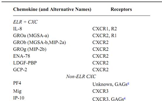
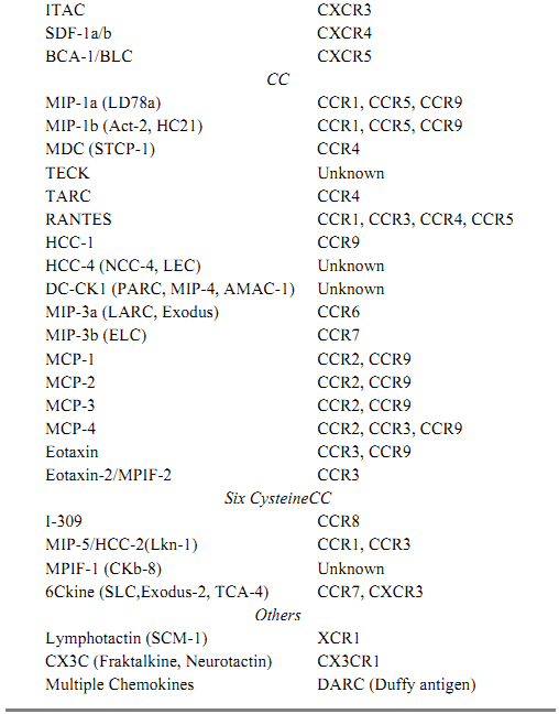
a GAG = Glycosyl amino glycan.
Members within a class are linked both by their structural similarities and by their location in the human genome. Although there are exceptions in each class, members of the CXC group generally map to the chromosomal locus 4q13. CC chemokines are found primarily on chromosomes 7, 8, 9, and 17. Chromosomal location may provide a useful tool in identifying new members of the g and d classes.
2. Chemokine Expression
Chemokines are expressed by virtually all cell types. Regulation of chemokine expression is complex and highly dependent on specific signaling pathways. Cells can express chemokines constitutively or in response to stimulation by, for example, interleukin-1 (IL-1), interleukin-4, tumor necrosis factor a, interferon-g (IFN-g), or pathogenic agents such as endotoxin (lipopolysaccharide). Low molecular-weight fragments (200 kDa) of the glycosaminoglycan hyaluronan (resulting from tissue destruction, for example) can enhance expression of some chemokines (eg, MIP-1, RANTES, or MCP-1) in specific macrophage types (eg, bone marrow-derived macrophages or elicited peritoneal macrophages) (7). Depending upon the situation, the expression of these same chemokines can be either suppressed or enhanced by inflammatory mediators, such as IL-10 or IFN-g (8).
One well-characterized example of induction of chemokines is provided by the macrophage. Macrophages can be stimulated to produce-chemokines during T cell-directed delayed type hypersensitive reactions in tissues by contact with activated T cells displaying CD40L. Kornbluth et al. (9) found that, although LPS was also a potent inducer of chemokines, the macrophage-stimulating lymphokines IFN-g and GM-CSF (granulocyte macrophage colony stimulating factor( produced by activated T cells were not good inducers.
3. Chemokine Receptors
Chemokines bind to seven-transmembrane-spanning G-protein-coupled receptors whose sequence homology is distinct from that of other such receptors. The interaction of a chemokine with its receptor can activate diverse signaling pathways, depending upon the specific signaling elements expressed by the target cell. Chemokine receptors are named on the basis of the cysteine motif that they recognize, and most will bind more than one chemokine ligand in that group. Likewise, a particular chemokine can interact with more than one receptor. Table 1 lists the chemokines and their respective receptors. Some receptors are expressed constitutively by particular cell types, whereas the expression of others may require an inducing signal. Thus CCR1 and CCR2 are constitutively expressed by monocytes, but are expressed by lymphocytes only after stimulation by IL-2 (10). Some chemokine receptors are expressed by nonhematopoietic cells, eg, endothelial cells, neurons, astrocytes, and epithelial cells. This emphasizes recent findings that chemokine receptors may have other important functions besides that of leukocyte chemotaxis. The Duffy antigen receptor, the determinant of the Duffy blood group, is found on a variety of cells, including erythrocytes and endothelial cells, as well as cells of the nervous system. It is unusual in its ability to bind both CC and CXC chemokines without eliciting any known biologic response. Possibly it serves to store or scavenge chemokines (11).
Specific chemokines generally act on specific subsets of leukocytes as a function of the receptors expressed by the cell (Table 2). Most CXC chemokines containing the ELR sequence are chemotactic for neutrophils, whereas non-ELR chemokines generally attract lymphocytes. Neutrophils express the CXCR1 receptor, which is stimulated by IL-8 and granulocyte chemotactic protein-2 (GCP-2), and the CXCR2 receptor, which responds to IL-8, GCP-2, the LPS-induced CXC chemokine (LIX), the neutrophil-activating peptide-2 (NAP-2), the epithelial cell-derived neutrophil activating peptide 78 (ENA-78), and the growth-regulated oncogenes, GRO-a, GRO-b and GRO-g. IL-2-stimulated lymphocytes express CXCR3, the receptor for MIG, IP-10, ITAC, and 6Ckine.
Table 2. Primary Expression of Chemokine Receptorsa
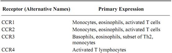
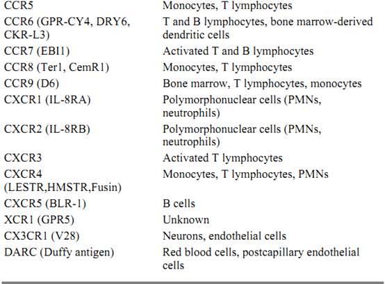
a This table lists cell types that have been widely accepted as expressors of chemokine receptors. It should be noted that many other cell types express these receptors as well.
Eosinophils, basophils, monocytes, NK cells, and T cells respond to members of the family of CC chemokines. These include monocyte chemoattractant proteins (MCP-1, -2, -3, -4, and -5), macrophage inflammatory protein-1a and -1b (MIP-1a, MIP-1b), RANTES (regulated on activation, normal T expressed and secreted), eotaxin-1 and -2, and I-309. Receptors for these chemokines expressed by eosinophils are CCR1 (recognized by MCP-3 and -4; MIP-1; RANTES) and CCR3 ) recognized by MCP-3 and -4; eotaxin-1 and -2; RANTES). Basophils express CCR3 and CCR2, which is activated by all the MCPs. Receptors expressed on monocytes are CCR1, 2, 5, and 8. Ligands for CCR5 include MIP-1a, MIP-1b, and RANTES, whereas the ligand for CCR8 is I-309. Activated T cells express CCR1, CCR2, CCR4 (recognized by TARC, the thymus and activation-regulated chemokine), CCR5, and CCR7, whose ligand is MIP-3 and 6Ckine (3, 4).
4. Chemokine Signaling
The ability of pertussis toxin to suppress many of the chemokine-induced signals suggests that the heterotrimeric G abg proteins mediating chemokine signaling belong to the Gi group, which is defined by their ability to inhibit adenylate cyclase. Activation of the Gi protein leads to replacement of GDP with GTP and dissociation into ai•GTP and bg subunits, both membrane-associated. Downstream mediators of general Gabg signaling may include the src of nonreceptor protein tyrosine kinases, phosphatidylinositol 3-kinase (PI3-K), phospholipase C, protein kinase C species, and the Ras-Raf-MEK-ERK kinase cascade (12, 13). Phospholipase C hydrolyzes phosphatidylinositol 4,5-bisphosphate to produce inositol trisphosphate, a mobilizer of intracellular Ca2+, and diacylglycerol, which, together with Ca2+, activates protein kinase C. Activation of phospholipase D, which generates phosphatidic acid, and phospholipase A2, which releases arachidonic acid, may be direct or indirect downstream effects of chemokine stimulation. Immediate effects (within seconds) on cell behavior are generally mediated by reversible changes in protein phosphorylation of cytoskeletal proteins, directed in part by the small Gtpases Rho, Rac and Cdc42, thereby affecting the polymerization and depolymerization of actin, which underlies the extension and retraction of lamellipodia, implementors of leukocyte migration (3). Longer term changes are effected by transcriptional or posttranscriptional changes in gene expression.
Evidence for specific signaling intermediates includes the following: The response of neutrophils to chemoattractants can be suppressed by inhibitors of tyrosine phosphorylation, possibly acting on members of the src of nonreceptor protein tyrosine kinases (12). In human T lymphocytes, RANTES induces PI3-K; also, wortmannin, which is a potent PI3-K inhibitor, can inhibit RANTES-induced T-lymphocyte responses (14). In a T-cell hybrid, MCP-1 mobilized intracellular Ca2+ concentrations, stimulated Ca2+ import, and transiently increased tyrosine phosphorylation of the ERKs by a pertussis toxin-sensitive process (15). In human monocytes, Yen et al. (16) obtained evidence for a putative Gq-mediated pathway, distinct from the pertussis toxin-sensitive Gi-dependent pathway, which activated ERK2 via a protein kinase C-dependent process that probably did not require Ras. Both the Gi and the proposed Gq pathways were required for a chemotactic response to MCP-1. MIP-1 activation of the CCR5 receptor expressed in a transfected murine pre-B lymphoma cell line led to phosphorylation and activation of a protein tyrosine kinase, known variously as RAFTK, Pyk2, or CAK-b, that is related to focal adhesion kinase (17). The cytoskeletal protein paxillin became phosphorylated and associated with RAFTK, and the downstream signaling kinases JNK/SAPK and p38 also appeared to be stimulated. Thus, chemokines have the ability to activate a variety of signal transduction mechanisms that connect the extracellular environment with the cytoskeleton.
5. Chemokine Functions
As part of the immune defense system, lymphocytes patrol the body, continually surveying it for pathogens or cells expressing abnormal surface antigens (11, 18). They move from the blood stream into tissues and then back into the blood stream via the lymphatic circulation. Granulocytes and monocytes exit the blood stream, but cannot recirculate. Extravasation is a coordinated multistep process that requires a series of complex changes in the activity of molecules on the cell's surface as it undergoes the transition from a freely floating cell to a weakly adherent rolling cell, and then to a strongly adherent stationary cell able to insinuate itself between the endothelial cells and penetrate the underlying stroma. Chemokines help drive this process and, as noted in Table 3, determine what types of cells infiltrate into the tissues in various inflammatory diseases. Imai et al. (19) report that the MDC and TARC chemokines, which are produced by dendritic cells in the thymus, act via the CCR4 receptor to recruit and activate T lymphocytes in the thymus. DC-CK1, another C-C chemokine derived from dendritic cells, is a chemoattractant for naive T cells (20). In allergic diseases such as asthma, expression of eotaxin and monocyte chemoattractant proteins appear particularly important in stimulating the accumulation and activation of eosinophils and mast cells, leading to histamine release and the allergic response (21). Similar events are thought to occur in rhinitis and atopic dermatitis.
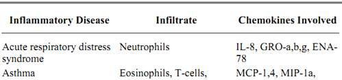
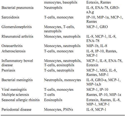
Table 3. Chemokine Involvement in Inflammatory Diseases
Subsets of lymphocytes express different surface molecules that allow them to target different tissues in the body. Extravasation of the lymphocyte from the blood stream is initiated when a free flowing lymphocyte becomes tethered to the endothelium such that it can still roll in the direction of flow. Rolling of leukocytes is controlled in part by the interaction of selectins with specific sialylated carbohydrate determinants. L-selectin, expressed by lymphocytes, monocytes, neutrophils, and eosinophils, interacts with receptors such as CD34 on the activated endothelium. P-selectin and E-selectin molecules are expressed by activated endothelial cells and bind structures related to the tetrasaccharide sialyl Lewis antigens on the leukocyte surface. Thrombin and histamine can mobilize P-selectin from intracellular stores, whereas up-regulation of E-selectin requires induction by inflammatory agents such as IL-1, tumor necrosis factor, or LPS. This interaction appears to result in activation of leukocyte 1 and 2 integrins, enabling them to bind to ligands of the immunoglobulin superfamily (eg, VCAM-1, the ICAMs; see Cell Adhesion Molecules) either expressed constitutively by the endothelial cells or induced by inflammatory agents. Various subsets of lymphocytes rolling on an ICAM-1 monolayer can be induced to arrest within one second by SDF-1, 6Ckine, MIP-3, or MIP-3; adhesion, which can be prevented by pertussis toxin, is transient, lasting some 5 to 8 minutes (22).
In inflammatory diseases, it is likely that sentinel cells at the site of inflammation produce chemokines that induce the strong interactions required to elicit leukocyte invasion. Chemokine expression has been demonstrated in glomerulonephritis, asthma, inflammatory bowel disease, and allogenic transplant rejection (2, 4). Because multiple inflammatory mediators are generally elaborated at sites of inflammation, the task of determining which is responsible for any particular pathogenic response is difficult. Although there is considerable redundancy built into the system, some chemokines predominate in some forms of injury. For example, IL-8 is a major causative factor associated with reperfusion injury and acid-induced pneumonitis, which is the consequence of neutrophil invasion and the release of inflammatory mediators (23).
Angiogenesis, the induction of synthesis of new blood vessels, typically with reference to the vascularization of a tumor, is a complex response affected by many factors, including chemokines. Although the specific role of chemokines in angiogenesis is unclear, evidence has suggested that CXC chemokines possessing the ELR motif generally promote angiogenesis, whereas those lacking the ELR motif tend to inhibit it (24). Mice lacking either SDF-1 or the counterpart receptor CXCR4 are defective in formation (vasculogenesis) of the large vessels supplying the intestinal tract (25). Vessels supplying the small intestine were missing major branches, and this led to hemorrhaging. Likewise, the vascular supply to the stomach in these mice was bereft of all large arteries and veins.
Many functions regulated by chemokines in the immune system, including cell migration and adhesion, as well as cytoskeletal reorganization, are important in the development and function of the nervous system. Defects in brain development in the CXCR4-deficient mouse (and its counterpart, the SDF-1 deficient mouse) are likely to be harbingers of other developmental and brain-specific functions of chemokines (26). The CXCR4-deficient mouse is embryonic-lethal, although with some animals surviving to term. The most prominent defects are found in the development of the cerebellum, where the granule neurons migrate prematurely. Since the radial glial cells are preserved in these animals, the evidence is consistent with an inhibitory role of SDF-1 on granule cell migration. This inhibition could occur by blocking the action of a migratory-promoting substance, or by altering adhesion molecules on the cell surface of the radial glial cell or the granule neuron. Since some of the guidance cues for this migratory process have been identified, it is of obvious importance to investigate if SDF-1 has any action to modulate the expression or conformation of these proteins.
Additional evidence for central nervous system actions of chemokines has been provided by the transgenic expression of chemokines under the influence of the myelin basic protein promoter. Directed expression of the murine chemokine KC stimulated neutrophil infiltration of the brain at various sites (27). Likewise, overexpression of MCP-1 in both thymus and brain led to infiltration of either MAC-1, F4/80, or macrophages and monocytes, respectively (28). These studies demonstrate that each organ is capable of recruiting mononuclear cells under specific conditions.
6. Viral Adaptation to Chemokines
Viruses often exploit endogenous cell surface receptors, including chemokine receptors, in the initial stages of infection. For example, mice deficient in MIP-1 as the result of a targeted gene disruption (knockout mice) exhibit attenuated responses to certain viral infections (Coxsackie virus, influenza virus). Several of the chemokine receptors serve as entry mechanisms for various strains of HIV. Roger (29) has reviewed the role of chemokine receptors and the HLA haplotype in the development of AIDS. The CCR5 receptor appears to be the preferred coreceptor for macrophage-tropic, nonsyncytium-forming HIV-1 strains, whereas the CXCR4 receptor (fusin) primarily services the syncytium-inducing, T-cell-line-tropic HIV-1 strains (30). Some HIV isolates can utilize both receptors, while a few other variants appear to have acquired the ability to co-opt other chemokine receptors as coreceptors for virus infection. Individuals with mutations in chemokine receptors may exhibit considerable resistance to infection or to progression of the disease. One mutation in particular should be noted. People homozygous for a deletion mutation in the CCR5 gene (CCR5 d32) are resistant to infection by the virus; heterozygotes for the same mutation had a significantly slower onset of disease (31). Chemokines can also affect the progression of an HIV infection. SDF-1, a ligand for CXCR4, inhibits HIV entry into cells (32, 33), and the CC chemokines MIP-1a, MIP-1b, and RANTES, all ligands for CCR5, likewise seem to retard HIV-1 infection (34).
7. Conclusion
The chemokine field is evolving rapidly. Although the initial focus has been on the role of chemokines in the immune system, future directions for chemokine research lie in elucidating chemokine expression and function in other organs and during development. Importantly, the involvement of chemokines and their receptors in many disease processes makes them primary targets for drug development.
References
1. N. W. Schluger and W. N. Rom (1997) Current Opin. Immunol. 9, 504–508.
2. B. J. Rollins (1997) Blood 90, 909–928.
3. M. Baggiolini (1998) Nature 392, 565–568.
4. A. D. Luster (1998) New England J. Med. 338, 436–445.
5. A. Zlotnick, J. Morales, and J. A. Hedrick (1998) Adv. Immunol. in press.
6. A. Pardigol et al. (1998) Proc. Natl. Acad. Sci. USA 95, 6308–6313.
7. C. McKee et al. (1996) J. Clin. Invest. 98, 2403–2413.
8. M. R. Horton et al. (1998) J. Immunol. 160, 3023–3030.
9. R. S. Kornbluth, K. Kee, and D. D. Richman (1998) Proc. Natl. Acad. Sci. USA 95, 5205–5210.
10. P. Loetscher, M. Seitz, M. Baggiolini, and B. Moser (1996) J. Exp. Med. 184, 569–577.
11. M. B. Furie and G. J. Randolph (1995) Amer. J. Pathol. 146, 1287–1301.
12. G. M. Bokoch (1995) Blood 86, 1649–1660.
13. J. S. Gutkind (1998) J. Biol. Chem. 273, 1839–1842.
14. L. Turner, S. G. Ward, and J. Westwick (1995) J. Immunol. 155, 2437–2444.
15. P. M. Dubois et al. (1996) J. Immunol. 156, 1356–1361.
16. H.-h. Yen, Y. Zhang, S. Penfold, and B. J. Rollins (1997) J. Leukoc. Biol. 61, 529–532.
17. R. K. Ganju et al. (1998) Blood 91, 791–797.
18. T. A. Springer (1994) Cell 76, 301–314.
19. T. Imai et al. (1998) J. Biol. Chem. 273, 1764–1768.
20. G. J. Adema et al. (1997) Nature 387, 713–717.
21. B. Lamkhioued et al. (1997) J. Immunol. 159, 4593–4601.
22. J. J. Campbell et al. (1998) Science 279, 381–384.
23. H. G. Folkesson, et al. (1995) J. Clin. Invest. 96, 107–116.
24. R. M. Strieter et al. (1995) J. Leukocyte Biol. 57, 752–762.
25. K. Tachinaba et al. (1998) Nature 393, 591–594.
26. Y-R. Zou et al. (1998) Nature 393, 595–599.
27. M. E. Fuentes et al. (1995) J. Immunol. 155, 5769–5776.
28. M. Tani et al. (1996) J. Clin. Invest. 98, 529–539.
29. M. Roger (1998) FASEB J. 12, 625–632.
30. Y. Feng, C. C. Broder, P. E. Kennedy, and E. A. Berger (1996) Science 272, 872–877.
31. M. W. Smith et al. (1997) Science 277, 959–965.
32. C. C. Bleul (1996) Nature 382, 829–833.
33. E. Oberlin et al. (1996) Nature 382, 833–835.
34. D. Zagury et al. (1998) Proc. Natl. Acad. Sci. USA 95, 3857–3861.


|
|
|
|
صنع الذكريات والتفكير يدمر الدماغ.. دراسة تشرح السبب
|
|
|
|
|
|
|
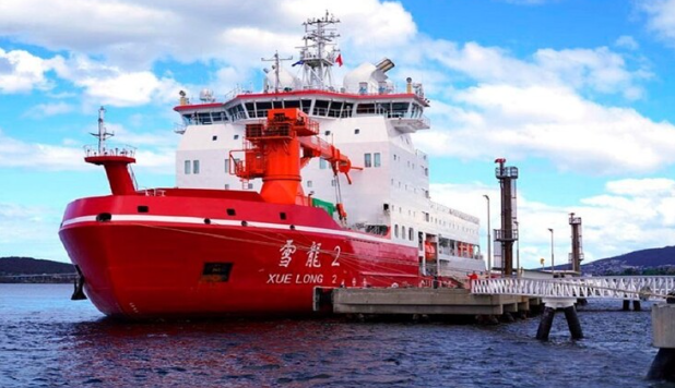
الصين.. عودة كاسحتي الجليد إلى شنغهاي بعد انتهاء بعثة استكشافية إلى القطب الجنوبي
|
|
|
|
|
|
جامعة الكفيل تكرم الفائزين بأبحاث طلبة كلية الصيدلة وطب الأسنان
|
|
|
|
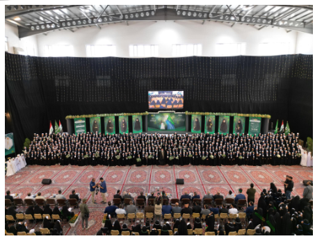
مشروع التكليف الشرعي بنسخته السادسة الورود الفاطمية... أضخم حفل لفتيات كربلاء
|
|
|
|
ضمن جناح جمعيّة العميد العلميّة والفكريّة المجمع العلمي يعرض إصداراته في معرض تونس الدولي للكتاب
|
|
|
|
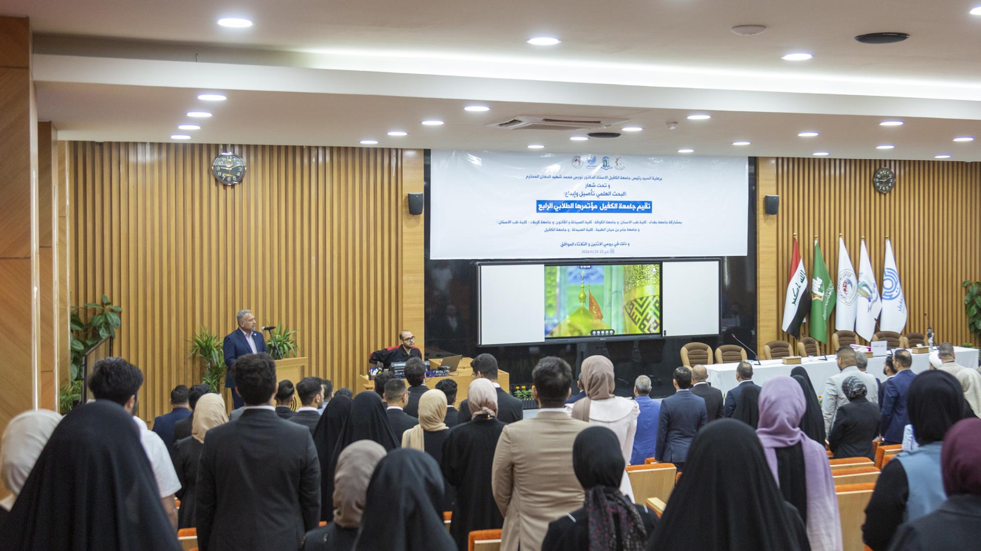
جامعة الكفيل تعقد مؤتمرها الطلابي العلمي الرابع
|